Arachnoid cyst
| Arachnoid cyst | |
|---|---|
 |
|
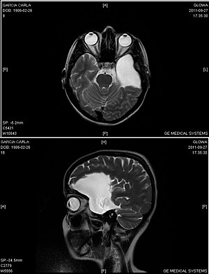
| An MRI of a 25-year-old woman with left frontotemporal arachnoid cyst. | |
| Classification and external resources | |
| Specialty | neurology |
| ICD-10 | G93.0 |
| ICD-9-CM | 348.0 |
| OMIM | 207790 |
| DiseasesDB | 33219 |
| eMedicine | radio/48 |
| MeSH | D016080 |
Arachnoid cysts are cerebrospinal fluid covered by arachnoidal cells and collagen that may develop between the surface of the brain and the cranial base or on the arachnoid membrane, one of the 3 meningeal layers that cover the brain and the spinal cord. Arachnoid cysts are a congenital disorder, and most cases begin during infancy; however, onset may be delayed until adolescence.
Arachnoid cysts can be found on the brain or on the spine. Intracranial arachnoid cysts usually occur adjacent to the arachnoidal cistern. Spinal arachnoid cysts may be extradural, intradural, or perineural and tend to present with signs and symptoms indicative of a radiculopathy.
Arachnoid cysts may also be classified as primary (congenital) or secondary (acquired) and have been reported in humans, cats, and dogs.
Arachnoid cysts can be relatively or present with symptoms; for this reason, diagnosis is often delayed.
Patients with arachnoid cysts may never show symptoms, even in some cases where the cyst is large. Therefore, while the presence of symptoms may provoke further clinical investigation, symptoms independent of further data cannot—and should not—be interpreted as evidence of a cyst's existence, size, location, or potential functional impact on the patient.
Symptoms vary by the size and location of the cyst(s), though small cysts usually have no symptoms and are discovered only incidentally. On the other hand, a number of symptoms may result from large cysts:
The exact cause of arachnoid cysts is not known. Researchers believe that most cases of arachnoid cysts are developmental malformations that arise from the unexplained splitting or tearing of the arachnoid membrane.
In some cases, arachnoid cysts occurring in the middle fossa are accompanied by underdevelopment (hypoplasia) or compression of the temporal lobe. The exact role that temporal lobe abnormalities play in the development of middle fossa arachnoid cysts is unknown.
There are some cases where hereditary disorders have been connected with arachnoid cysts.
Some complications of arachnoid cysts can occur when a cyst is damaged because of minor head trauma. Trauma can cause the fluid within a cyst to leak into other areas (e.g., subarachnoid space). Blood vessels on the surface of a cyst may tear and bleed into the cyst (intracystic hemorrhage), increasing its size. If a blood vessel bleeds on the outside of a cyst, a collection of blood (hematoma) may result. In the cases of intracystic hemorrhage and hematoma, the individual may have symptoms of increased pressure within the cranium and signs of compression of nearby nerve (neural) tissue.
...
Wikipedia
