Diffusion MRI
| Diffusion MRI | |
|---|---|
| Medical diagnostics | |

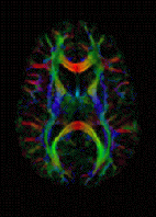
DTI Color Map
|
|
| MeSH | D038524 |
Diffusion-weighted magnetic resonance imaging (DWI or DW-MRI) is an imaging method that uses the diffusion of water molecules to generate contrast in MR images. It allows the mapping of the diffusion process of molecules, mainly water, in biological tissues, in vivo and non-invasively. Molecular diffusion in tissues is not free, but reflects interactions with many obstacles, such as macromolecules, fibers, and membranes. Water molecule diffusion patterns can therefore reveal microscopic details about tissue architecture, either normal or in a diseased state. A special kind of DWI, diffusion tensor imaging (DTI), has been used extensively to map white matter tractography in the brain.
The first diffusion MRI images of the normal and diseased brain were made public in 1985. Its main clinical application has been in the study and treatment of neurological disorders, especially for the management of patients with acute stroke. Because it can reveal abnormalities in white matter fiber structure and provide models of brain connectivity, it is rapidly becoming a standard for white matter disorders. The ability to visualize anatomical connections between different parts of the brain, noninvasively and on an individual basis, has emerged as a major breakthrough for neuroscience's Human Connectome Project. More recently, a new field has emerged, diffusion functional MRI (DfMRI) as it was suggested that with DWI one could also get images of neuronal activation in the brain. Finally, the method of diffusion MRI has also been shown to be sensitive to perfusion, as the movement of water in blood vessels mimics a random process, intravoxel incoherent motion (IVIM). IVIM dMRI is rapidly becoming a major method to obtain images of perfusion in the body, especially for cancer detection and monitoring.
...
Wikipedia
