Hepatic adenoma
| Hepatocellular adenoma | |
|---|---|
 |
|
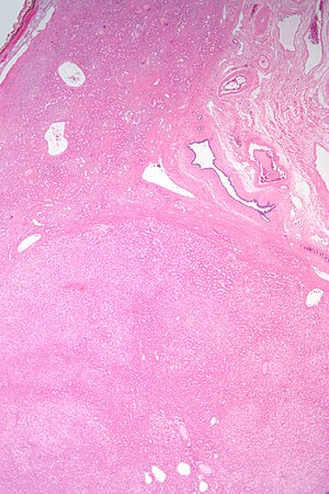
| Micrograph of a hepatic adenoma (bottom of image). H&E stain | |
| Classification and external resources | |
| ICD-O | M8170/0 |
| DiseasesDB | 5726 |
| eMedicine | med/48 |
| MeSH | D018248 |
Hepatocellular adenoma, also hepatic adenoma, or rarely hepadenoma, is an uncommon benign liver tumor which is associated with increased levels of estrogen. Patients of advanced age, or taking higher potency hormones, or patients with prolonged duration of use have a significantly increased risk of developing hepatocellular adenomas.
About 25–50% of hepatic adenomas cause pain in the right upper quadrant or epigastric region of the abdomen. Since hepatic adenomas can be large (8–15 cm), patients may notice a palpable mass. However, hepatic adenomas are usually asymptomatic, and may be discovered incidentally on imaging ordered for some unrelated reason. If not treated, there is a 30% risk of bleeding. Bleeding may lead to hypotension, tachycardia, and sweating (diaphoresis).
Ninety percent of hepatic adenomas arise in women aged 20–40, most of whom use oral contraceptives.
It is important to distinguish hepatic adenoma from other benign liver tumors, such as hemangiomas and focal nodular hyperplasia, because hepatic adenomas have a small but meaningful risk of progressing into a malignancy.MRI is the most useful investigation in the diagnosis and work-up. A poly-phasic CT scan is another useful test for diagnosing hepatic adenoma.
Large hepatic adenomas have a tendency to rupture and bleed massively inside the abdomen.
Hepatic adenomas are, typically, well-circumscribed nodules that consist of sheets of with a bubbly vacuolated cytoplasm. The hepatocytes are on a regular reticulin scaffold and less or equal to three cell thick.
The histologic diagnosis of hepatic adenomas can be aided by reticulin staining. In hepatic adenomas, the reticulin scaffold is preserved and hepatocytes do not form layers of four or more hepatocytes, as is seen in .
Cells resemble normal hepatocytes and are traversed by blood vessels but lack portal tracts or central veins.
Micrograph of hepatic adenoma. H&E stain
...
Wikipedia
