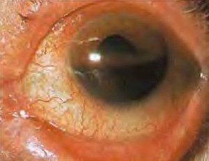
Hyphema
| Hyphema | |
|---|---|
 |
|
| Hyphema - occupying half of anterior chamber of eye | |
| Classification and external resources | |
| Specialty | ophthalmology |
| ICD-10 | H21.0 |
| ICD-9-CM | 364.41 |
| DiseasesDB | 31299 |
| MedlinePlus | 001021 |
| eMedicine | oph/765 |
| MeSH | C11.290.484 |
Hyphema (or hyphaema, see spelling differences) is blood in the front (anterior) chamber of the eye. It may appear as a reddish tinge, or it may appear as a small pool of blood at the bottom of the iris or in the cornea.
As of 2012, the rate of hyphemas in the United States are about 20 cases per 100,000 peopleTemplate:Specify inline annually.
Hyphemas are frequently caused by injury, and may partially or completely block vision. The most common causes of hyphema are intraocular surgery, blunt trauma, and lacerating trauma. Hyphemas may also occur spontaneously, without any inciting trauma. Spontaneous hyphemas are usually caused by the abnormal growth of blood vessels (neovascularization), tumors of the eye (retinoblastoma or iris melanoma), uveitis, or vascular anomalies (juvenile xanthogranuloma). Additional causes of spontaneous hyphema include: rubeosis iridis, myotonic dystrophy, leukemia, hemophilia, and von Willebrand disease. Conditions or medications that cause thinning of the blood, such as aspirin, warfarin, or drinking alcohol may also cause hyphema.
Hyphemas require urgent assessment by an optometrist or ophthalmologist as they may result in permanent visual impairment.
A long-standing hyphema may result in hemosiderosis and heterochromia. Blood accumulation may also cause an elevation of the intraocular pressure. On average, the increased pressure in the eye remains for six days before dropping. Most uncomplicated hyphemas resolve within 5–6 days.
...
Wikipedia
