Iliopsoas group
| Iliopsoas | |
|---|---|

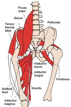
Anterior hip muscles. The iliopsoas is not labeled but can be seen as the psoas major and the iliacus join inferiorly.
|
|
| Details | |
| Origin | Iliac fossa and lumbar spine |
| Insertion | Lesser trochanter of femur |
| Artery | Medial femoral circumflex artery and iliolumbar artery |
| Nerve | Branches from L1 to L3 |
| Actions | Flexion of hip |
| Antagonist | Gluteus maximus and the posterior compartment of thigh |
| Identifiers | |
| Latin | Musculus iliopsoas |
| Dorlands /Elsevier |
m_22/12549330 |
| TA | A04.7.02.002 |
| FMA | 64918 |
|
Anatomical terms of muscle
[]
|
|
The iliopsoas (ilio-so-as) refers to the joined psoas and the iliacus muscles. The two muscles are separate in the abdomen, but usually merge in the thigh. As such, they are usually given the common name iliopsoas. The iliopsoas muscle joins to the femur at the lesser trochanter, and acts as the strongest of the hip.
The iliopsoas muscle is supplied by the lumbar spinal nerves L1-3 (psoas) and parts of the femoral nerve (iliacus).
The iliopsoas muscle is a composite muscle formed from the psoas major muscle, and the iliacus muscle. The psoas major originates along the outer surfaces of the vertebral bodies of T12 and L1-L3 and their associated intervertebral discs. The iliacus originates in the iliac fossa of the pelvis.
The psoas major unites with the iliacus at the level of the inguinal ligament and crosses the hip joint to insert on the lesser trochanter of the femur. The iliopsoas is classified as the "dorsal hip muscles". or "inner hip muscles". The psoas minor does contribute to the iliopsoas muscle.
The psoas major is innervated by direct branches of the anterior rami off the lumbar plexus at the levels of L1-L3, while the iliacus is innervated by the femoral nerve (which is composed of nerves from the anterior rami of L2-L4).
...
Wikipedia
