Myocyte
| Myocyte | |
|---|---|

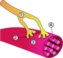
General structure of a muscle cell and neuromuscular junction:
|
|
| Details | |
| Identifiers | |
| Latin | Myocytus |
| Code | TH H2.00.05 |
|
Anatomical terminology
[]
|
|
A myocyte (also known as a muscle cell) is the type of cell found in muscle tissue. Myocytes are long, tubular cells that develop from myoblasts to form muscles in a process known as myogenesis. There are various specialized forms of myocytes: cardiac, skeletal, and smooth muscle cells, with various properties. The striated cells of cardiac and skeletal muscles are referred to as muscle fibers.Cardiomyocytes are the muscle fibres that form the chambers of the heart, and have a single central nucleus. Skeletal muscle fibers help support and move the body and tend to have peripheral nuclei. Smooth muscle cells control involuntary movements such as the peristalsis contractions in the oesophagus and stomach.
The unusual microstructure of muscle cells has led cell biologists to create specialized terminology. However, each term specific to muscle cells has a counterpart that is used in the terminology applied to other types of cells:
The sarcoplasm is the cytoplasm of a muscle fiber. Most of the sarcoplasm is filled with myofibrils, which are long protein cords composed of myofilaments. The sarcoplasm is also composed of glycogen, a polysaccharide of glucose monomers, which provides energy to the cell with heightened exercise, and myoglobin, the red pigment that stores oxygen until needed for muscular activity.
There are three types of myofilaments:
Together, these myofilaments work to produce a muscle contraction.
The sarcoplasmic reticulum, a specialized type of smooth endoplasmic reticulum, forms a network around each myofibril of the muscle fiber. This network is composed of groupings of two dilated end-sacs called terminal cisternae, and a single transverse tubule, or T tubule, which bores through the cell and emerge on the other side; together these three components form the triads that exist within the network of the sarcoplasmic reticulum, in which each T tubule has two terminal cisternae on each side of it. The sarcoplasmic reticulum serves as reservoir for calcium ions, so when an action potential spreads over the T tubule, it signals the sarcoplasmic reticulum to release calcium ions from the gated membrane channels to stimulate a muscle contraction.
...
Wikipedia
