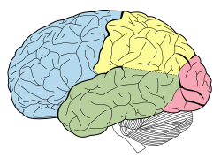
Occipital cortex
| Occipital lobe | |
|---|---|
|
Lobes of the human brain (the occipital lobe is shown in red)
|
|

|
|
| Details | |
| Part of | cerebrum |
| Artery | posterior cerebral artery |
| Identifiers | |
| Latin | lobus occipitalis |
| MeSH | A08.186.211.730.885.213.571 |
| NeuroNames | hier-122 |
| NeuroLex ID | Occipital lobe |
| TA | A14.1.09.132 |
| FMA | 67325 |
|
Anatomical terms of neuroanatomy
[]
|
|
The occipital lobe is one of the four major lobes of the cerebral cortex in the brain of mammals. The occipital lobe is the visual processing center of the mammalian brain containing most of the anatomical region of the visual cortex. The primary visual cortex is Brodmann area 17, commonly called V1 (visual one). Human V1 is located on the medial side of the occipital lobe within the calcarine sulcus; the full extent of V1 often continues onto the posterior pole of the occipital lobe. V1 is often also called striate cortex because it can be identified by a large stripe of myelin, the Stria of Gennari. Visually driven regions outside V1 are called extrastriate cortex. There are many extrastriate regions, and these are specialized for different visual tasks, such as visuospatial processing, color differentiation, and motion perception. The name derives from the overlying occipital bone, which is named from the Latin ob, behind, and caput, the head. Bilateral lesions of the occipital lobe can lead to cortical blindness (See Anton's syndrome).
The two occipital lobes are the smallest of four paired lobes in the human cerebral cortex. Located in the rearmost portion of the skull, the occipital lobes are part of the forebrain. None of the cortical lobes are defined by any internal structural features, but rather by the bones of the head bone that overlie them. Thus, the occipital lobe is defined as the part of the cerebral cortex that lies underneath the occipital bone. (See the human brain article for more information.)
...
Wikipedia

